
 英语
英语 西班牙语
西班牙语 德语
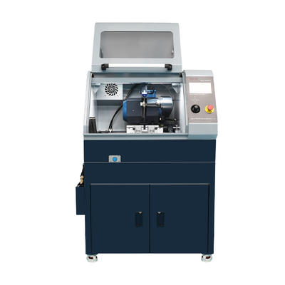
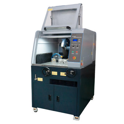
德语Beta-300Pro Automatic Abrasive Sectioning Machine
Beta-300Pro is a fully automatic vertical metallographic cutting machine. It utilizes advanced intel...
We use first- and third-party cookies including other tracking technologies from third party publishers to give you the full functionality of our website, to customize your user experience, perform analytics and deliver personalized advertising on our websites, apps and newsletters across internet and via social media platforms. For that purpose, we collect information about user, browsing patterns and device.
By clicking "Accept All Cookies", you accept this, and agree that we share this information with third parties, such as our advertising partners. If you prefer, you can choose to continue with "Only Required Cookies". But keep in mind that blocking some types of cookies may impact how we can deliver tailored content that you might like.
For more information and to customize your options, click on "Cookie settings". If you want to learn more about cookies and why we use them, visit our Cookie Policy page at any time. Cookie Policy

Material failure investigations, quality control checkpoints, and research labs all rely on one instrument category that has not been replaced by newer technologies: the optical microscope. While electron microscopy offers extreme magnification, most day-to-day microscopy & optical analysis work is still performed on the bench, where speed, sample preparation simplicity, and cost make optical systems the practical first choice.
The challenge for labs and production floors is not whether to use a microscope, but which configuration actually matches the material being examined. Grain structure evaluation, coating thickness checks, weld inspection, and fracture surface review each demand different combinations of resolution, contrast method, and stage design. Choosing incorrectly leads to underused equipment, slower throughput, and inconsistent reporting.
Before comparing specific instruments, it helps to place metallurgical systems within the broader landscape of types of microscopes used in industrial and laboratory settings. Each design solves a different observation problem.
| Microscope Type | Primary Use Case | Typical Sample |
|---|---|---|
| Stereo Microscope | Low-power surface inspection | Assemblies, castings |
| Metallurgical Microscope | Reflected light structure analysis | Polished metal sections |
| Digital Microscope | Live imaging and measurement | Mixed materials |
| Polarizing Microscope | Birefringence and phase identification | Minerals, ceramics |
| Confocal Microscope | 3D surface reconstruction | Fine machined surfaces |
Of these, the metallurgical category is the workhorse for metals and coatings inspection because it is built specifically around opaque, non-transparent samples viewed with reflected rather than transmitted light.
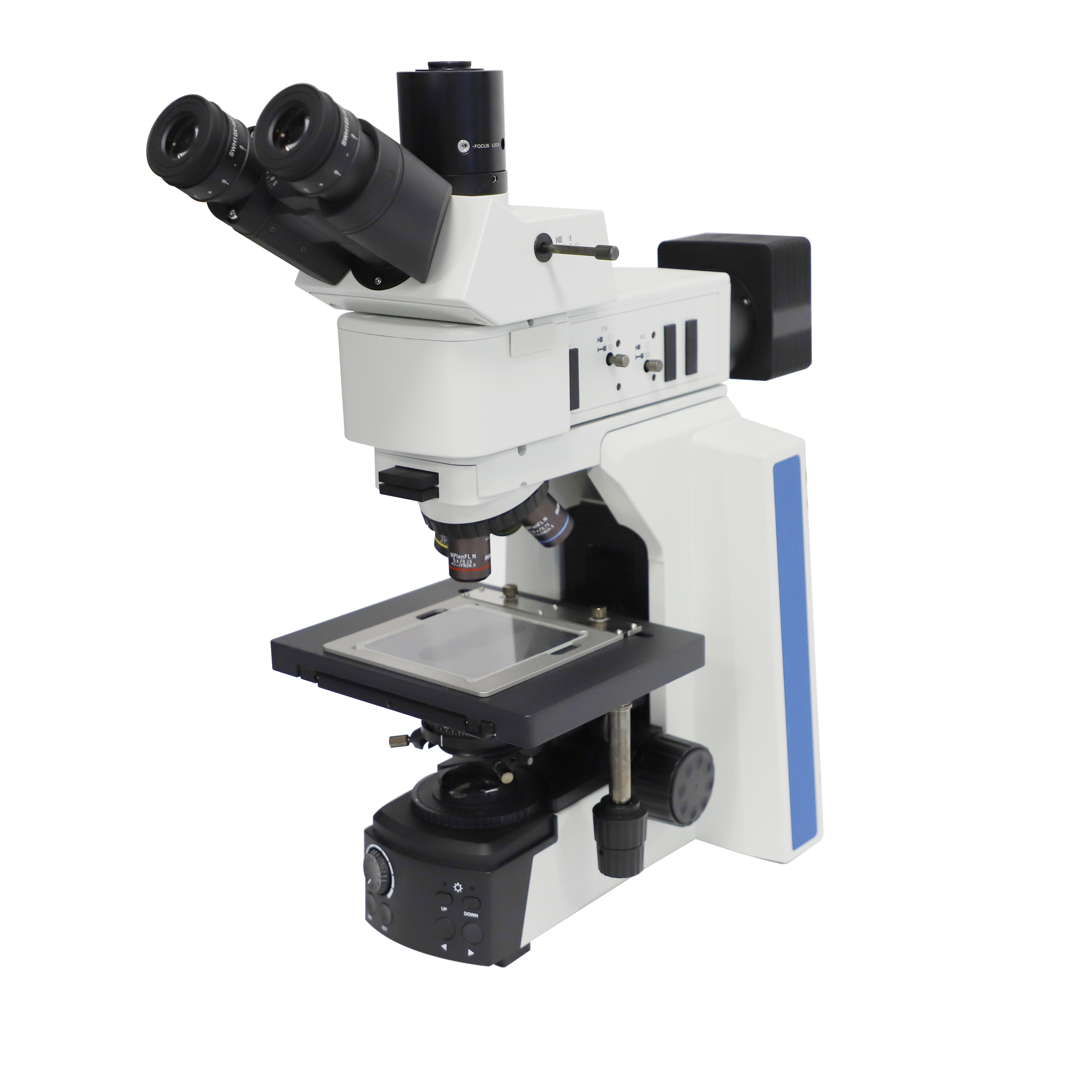
Unlike biological microscopes that pass light through a thin, transparent slide, metallurgical microscopes illuminate the sample from above and capture the light reflected off a polished, often etched, surface. This reflected-light design is essential because metals, ceramics, and many composites cannot be sliced thin enough for light to pass through without destroying the very structure being studied.
Three structural elements separate a metallurgical system from a general-purpose microscope:
Grain boundary visibility, inclusion counting, and case-depth measurement all depend on the quality of this reflected-light path, not just the magnification number printed on the objective.
Buyers frequently compare microscopes by magnification alone, but magnification without adequate resolving power simply produces a larger, blurrier image. Resolution is governed by the numerical aperture of the objective lens and the wavelength of the illumination, not by the eyepiece.
Two objective lenses with identical magnification but different numerical apertures will show visibly different levels of detail on the same grain structure. This is why lens selection, not magnification selection, should drive purchasing decisions for demanding structural analysis.
| Numerical Aperture | Approx. Resolving Power | Suitable Application |
|---|---|---|
| 0.25 | Coarse detail | General surface scan |
| 0.65 | Medium detail | Grain size estimation |
| 0.90 and above | Fine detail | Inclusion rating, fine porosity |
Polished metal surfaces often show very little contrast under plain bright field illumination, especially before etching. Several optical techniques increase contrast without altering the sample chemically.
Selecting a system with built-in switching between these modes saves significant time compared to instruments where filters and analyzers must be manually installed for each observation type.
Specification sheets rarely tell the whole story. The following factors are the ones that most affect daily usability in an industrial or laboratory environment.
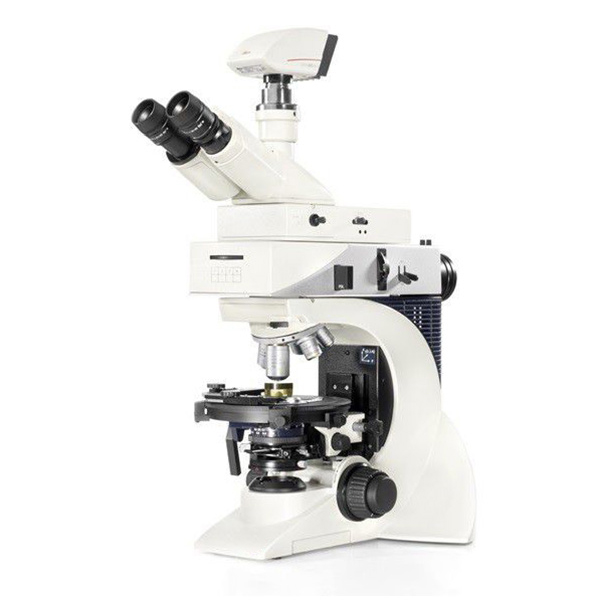
Within reflected-light instrumentation, leica metallographic microscopes are frequently specified in laboratories that require both routine grain analysis and periodic polarized or DIC observation on the same bench. Their modular illuminator housings allow a technician to move between bright field, dark field, and polarized modes without reconfiguring the optical path each time.
For labs standardizing across multiple inspection stations, consistency of image color rendering and stage repeatability matters as much as raw resolution. Instruments in this category are commonly paired with digital capture systems so that grain size ratings, inclusion counts, and coating thickness measurements can be recorded against a documented image rather than a visual estimate alone.
| Inspection Task | Recommended Contrast Mode | Typical Magnification |
|---|---|---|
| Grain size rating | Bright field, etched surface | 100x to 400x |
| Inclusion counting | Bright field, unetched surface | 100x to 500x |
| Coating thickness | Bright field, cross-section | 200x to 1000x |
| Weld structure review | Polarized or dark field | 50x to 200x |
Rather than starting from a product catalog, it is more effective to define the inspection task first and work backward to the required specification.
Following this sequence prevents the common mistake of purchasing based on magnification headline numbers while overlooking illumination flexibility, which is usually the more limiting factor in daily operation.
A metallurgical microscope uses reflected light directed down through the objective onto an opaque sample, while a standard biological microscope passes transmitted light through a thin, translucent slide. This difference makes metallurgical systems suitable for metals, ceramics, and other non-transparent materials.
Most grain size evaluations are performed between 100x and 400x. Higher magnification is reserved for fine inclusion counting or detailed coating cross-sections, where features are smaller than typical grain boundaries.
Numerical aperture controls how much fine detail the objective can resolve. A high magnification objective with low numerical aperture will enlarge the image without revealing additional structural detail, producing a soft or blurry result.
Polarized light is not required for all inspection tasks, but it becomes essential when examining grain orientation, certain ceramic phases, or materials with birefringent properties that are invisible under standard bright field illumination.
A well-specified system with interchangeable objectives, multiple contrast modes, and a stable stage can cover both routine quality checks and more detailed structural research, reducing the need for separate dedicated instruments in many facilities.

.png?imageView2/2/w/400/format/jpg/q/75 "Disposable Sample Cups")

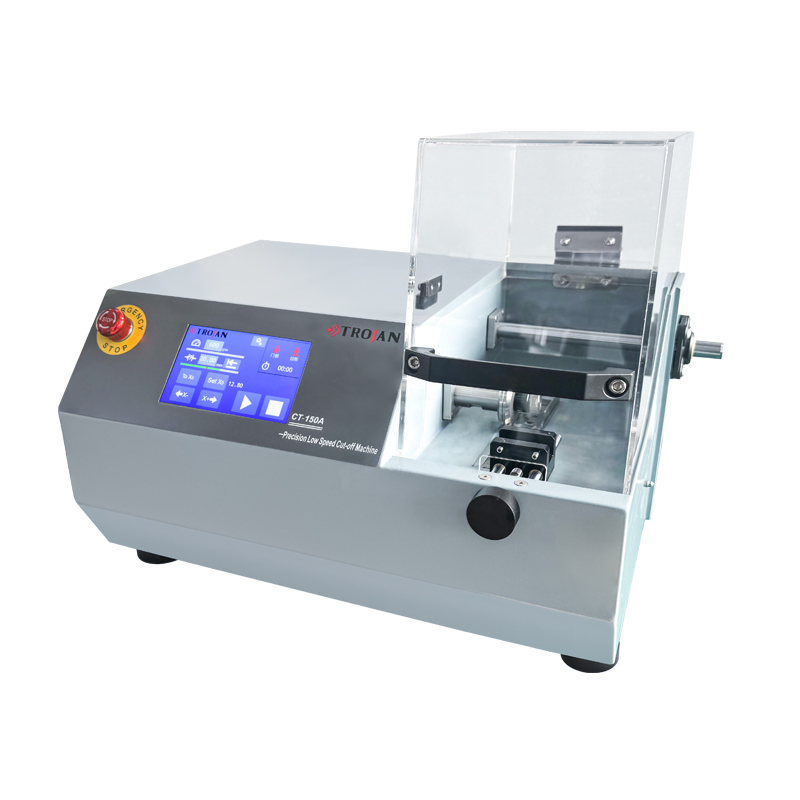


Based in China, TROJAN pays attention to the international market, and has sold metallographic equipment to more than 50 countries, such as Europe, America, Japan and South Korea, Southeast Asia countries and regions


